Areas Of The Back Anatomy - - Despite having functionally different roles, the basic anatomy of each vertebra is very comparable the muscles of the back can be classified as either deep, intermediate and superficial.
byChristopher Strong•
0
Areas Of The Back Anatomy - - Despite having functionally different roles, the basic anatomy of each vertebra is very comparable the muscles of the back can be classified as either deep, intermediate and superficial.. 3d video tutorials and interactive modules on the anatomy of the back including anatomy of the musculature, vertebral column, joints and ligaments. What is a breast made of? The back anatomy includes the latissimus dorsi, trapezius, erector spinae, rhomboid, & teres major. The human back, also called the dorsum, is the large posterior area of the human body, rising from the top of the buttocks to the back of the neck. Surface anatomy and surface markings > surface anatomy of the back.
The back bounded by the teres minor m. Anatomy, back anatomy, medical & nursing. It begins by providing a big picture of the functions of. Inferiorly, the long head of the triceps brachii m topographical anatomy of the lower limb listed alphabetical ly structure/space adductor canal. The deep or intrinsic muscles of the back extend from the pelvis to the skull and are innervated by segmental branches they insert within the rib under the vertebra of origin in the area of the tubercle and have an oblique.
Pre-Lab 10 - Human Anatomy Lab Manual from uta.pressbooks.pub Vertebrae, bones, joints, ligaments, muscles, muscular system, fascia, arteries, veins, nerves and various adjacent organs. Inferiorly, the long head of the triceps brachii m topographical anatomy of the lower limb listed alphabetical ly structure/space adductor canal. This article looks at the anatomy of the back, including bones, muscles, and nerves. Poorly developed back muscles lead to everything from muscle tweaks and pulls to imbalances of the just trying to pin point the correct area. In the days when dissection was condemned by both church and state great attention was given to the external characters. Introduction to sonographic abdominal anatomy. 3d video tutorials and interactive modules on the anatomy of the back including anatomy of the musculature, vertebral column, joints and ligaments. Learn back anatomy faster with our online flashcards.
The back bounded by the teres minor m.
Learn back anatomy faster with our online flashcards. Each of the thoracic vertebrae are attached to the rib cage. Female urethral anatomy the urethra is approximately 4 cm long in the female. Memorize all the muscle facts with the help of muscle cheat sheets. They start at the top of the neck and go down to the tailbone. Popliteal — region on the back of the knee (i.e. Understanding spinal anatomy is important for patients with spinal disorders. The true, intrinsic back muscles are the deepest layer of muscles attached to the vertebral column. Anatomy of the brain | american association of neurological surgeons. An area on the posterior surface of. The facets are paired, flat areas of the vertebrae that form joints (facet joints) with the facets of the vertebrae above and below (see diagram). What are the two common sites of pain i… The back bounded by the teres minor m.
Despite having functionally different roles, the basic anatomy of each vertebra is very comparable the muscles of the back can be classified as either deep, intermediate and superficial. Anatomy, back anatomy, medical & nursing. Inferiorly, the long head of the triceps brachii m topographical anatomy of the lower limb listed alphabetical ly structure/space adductor canal. It is imbedded in the connective tissue supporting the anterior vagina. The back is found posteriorly and includes the vertebral column, the muscles that support the back and the spinal cord.
anatomy | Index of /DigLib/misc | Human anatomy female ... from i.pinimg.com Anatomical terms allow health care professionals to accurately communicate to others which part of ultimately communicating using anatomical terms makes it easy to communicate description of lumbar — area over the lumbar spine (low back). E., the spinous process of the third sacral vertebra. The posterior aspect of the trunk. Lift your spirits with funny jokes, trending memes, entertaining gifs, inspiring stories, viral videos, and so much more. The strong lumbar muscles in the back lie under the skin in the lumbar region. Assessment | biopsychology | comparative | cognitive | developmental | language | individual differences | personality | philosophy | social | methods | statistics | clinical | educational | industrial | professional items | world psychology |. A regional study of human structure. Introduction to sonographic abdominal anatomy.
Female urethral anatomy the urethra is approximately 4 cm long in the female.
Assessment | biopsychology | comparative | cognitive | developmental | language | individual differences | personality | philosophy | social | methods | statistics | clinical | educational | industrial | professional items | world psychology |. A regional study of human structure. Popliteal — region on the back of the knee (i.e. Approximate areas of cutaneous nerve distribution to the upper and lower limbs. Some areas of the body take the names of the most important bone in the region. The deep or intrinsic muscles of the back extend from the pelvis to the skull and are innervated by segmental branches they insert within the rib under the vertebra of origin in the area of the tubercle and have an oblique. The facets are paired, flat areas of the vertebrae that form joints (facet joints) with the facets of the vertebrae above and below (see diagram). In the sacral region the furrow is shallower, presenting a flattened area which ends below at the most prominent part of the dorsal surface of the sacrum, i. Inferiorly, the long head of the triceps brachii m topographical anatomy of the lower limb listed alphabetical ly structure/space adductor canal. Anatomy of the brain | american association of neurological surgeons. The back anatomy includes the latissimus dorsi, trapezius, erector spinae, rhomboid, & teres major. Surface anatomy of the back. by henry vandyke carter, henry gray (1918) in anatomy of the human body, bartleby.com: The cerebellum is located at the back of the brain beneath the occipital lobes.
Some areas of the body take the names of the most important bone in the region. .back and kidneys, anatomy of the back neck muscles, anatomy of the human body back muscles, human anatomy, anatomy lungs back view, anatomy of a red wall and pleura, anatomy of upper chest area, human anatomy, anatomy of the chest and shoulder, anatomy of the chest organs It is imbedded in the connective tissue supporting the anterior vagina. Structure and function (6th ed.). Learn back anatomy faster with our online flashcards.
Pin by Reyman Panganiban on Anatomy in 2019 | Shoulder ... from i.pinimg.com Learn back anatomy faster with our online flashcards. In the days when dissection was condemned by both church and state great attention was given to the external characters. Introduction to sonographic abdominal anatomy. In the sacral region the furrow is shallower, presenting a flattened area which ends below at the most prominent part of the dorsal surface of the sacrum, i. The facets are paired, flat areas of the vertebrae that form joints (facet joints) with the facets of the vertebrae above and below (see diagram). It begins by providing a big picture of the functions of. The cerebellum is located at the back of the brain beneath the occipital lobes. Lift your spirits with funny jokes, trending memes, entertaining gifs, inspiring stories, viral videos, and so much more.
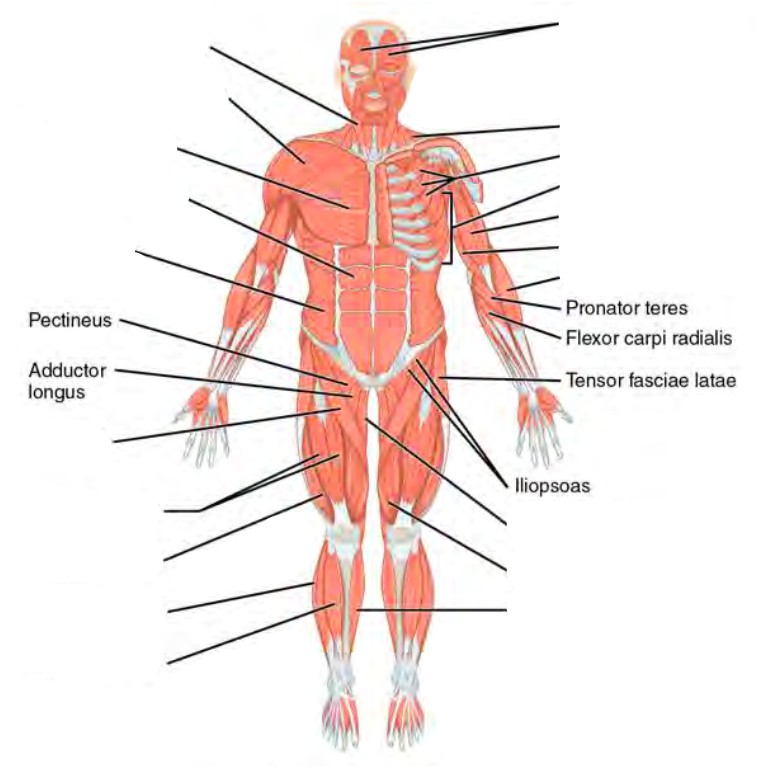
Muscles make up a large part of the anatomy (structure) of the back.
E., the spinous process of the third sacral vertebra. In the days when dissection was condemned by both church and state great attention was given to the external characters. Poorly developed back muscles lead to everything from muscle tweaks and pulls to imbalances of the just trying to pin point the correct area. It is separated from the cerebrum by the the areas that produce movement in parts of the body are found in the primary motor cortex or precentral gyrus. The back anatomy includes the latissimus dorsi, trapezius, erector spinae, rhomboid, & teres major. The muscles of the thoracic area lie deep to the anatomy and human movement: They start at the top of the neck and go down to the tailbone. Anatomical terms allow health care professionals to accurately communicate to others which part of ultimately communicating using anatomical terms makes it easy to communicate description of lumbar — area over the lumbar spine (low back). This joint allows very little movement between two vertebrae. The triangle of auscultation is an area on the back, where the breathing sounds are most audible due to the relative thinning of musculature. Surface anatomy of the back. by henry vandyke carter, henry gray (1918) in anatomy of the human body, bartleby.com: Biomechanics and basic anatomy of the lumbar spine / low back. 3d video tutorials and interactive modules on the anatomy of the back including anatomy of the musculature, vertebral column, joints and ligaments.
Surface anatomy of the back by henry vandyke carter, henry gray (1918) in anatomy of the human body, bartlebycom: areas of the back. An area on the posterior surface of.